What Is Genetic Code and How Is It Read

The ribosome assembles the polypeptide chain
To manufacture protein molecules, a jail cell must kickoff transfer information from DNA to mRNA through the procedure of transcription. Then, a process chosen translation uses this mRNA as a template for protein assembly. In fact, this menstruum of data from DNA to RNA and finally to protein is considered the cardinal dogma of genetics, and it is the starting signal for understanding the function of the genetic information in Deoxyribonucleic acid.
Only merely how does translation work? In other words, how does the prison cell read and translate the information that is stored in DNA and carried in mRNA? The answer to this question lies in a serial of circuitous mechanisms, almost of which are associated with the cellular structure known as the ribosome. In order to sympathize these mechanisms, however, it's kickoff necessary to take a closer await at the concept known as the genetic code.
What is the genetic code?
At its heart, the genetic lawmaking is the fix of "rules" that a cell uses to interpret the nucleotide sequence inside a molecule of mRNA. This sequence is broken into a serial of three-nucleotide units known as codons (Figure 1). The three-letter nature of codons means that the four nucleotides plant in mRNA — A, U, G, and C — can produce a total of 64 different combinations. Of these 64 codons, 61 stand for amino acids, and the remaining three represent finish signals, which trigger the end of protein synthesis. Because at that place are merely 20 different amino acids simply 64 possible codons, most amino acids are indicated by more than than one codon. (Annotation, however, that each codon represents simply 1 amino acid or end codon.) This miracle is known as back-up or degeneracy, and it is important to the genetic code because it minimizes the harmful furnishings that incorrectly placed nucleotides can take on poly peptide synthesis. Yet some other cistron that helps mitigate these potentially damaging furnishings is the fact that there is no overlap in the genetic code. This means that the three nucleotides within a item codon are a part of that codon merely — thus, they are non included in either of the adjacent codons.
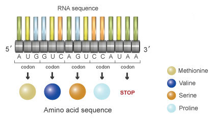
Figure 1: In mRNA, three-nucleotide units called codons dictate a particular amino acid. For example, AUG codes for the amino acrid methionine (beige).
The idea of codons was first proposed by Francis Crick and his colleagues in 1961. During that same twelvemonth, Marshall Nirenberg and Heinrich Matthaei began deciphering the genetic lawmaking, and they determined that the codon UUU specifically represented the amino acid phenylalanine. Post-obit this discovery, Nirenberg, Philip Leder, and Har Gobind Khorana eventually identified the residual of the genetic code and fully described which codons corresponded to which amino acids.
Reading the genetic code
Redundancy in the genetic lawmaking means that most amino acids are specified by more than than ane mRNA codon. For example, the amino acid phenylalanine (Phe) is specified by the codons UUU and UUC, and the amino acid leucine (Leu) is specified past the codons CUU, CUC, CUA, and CUG. Methionine is specified by the codon AUG, which is also known as the starting time codon. Consequently, methionine is the first amino acid to dock in the ribosome during the synthesis of proteins. Tryptophan is unique because it is the only amino acid specified by a single codon. The remaining 19 amino acids are specified by between two and six codons each. The codons UAA, UAG, and UGA are the terminate codons that point the termination of translation. Figure two shows the 64 codon combinations and the amino acids or stop signals they specify.
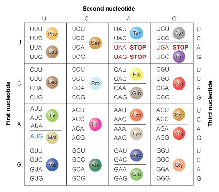
Figure ii: The amino acids specified by each mRNA codon. Multiple codons tin lawmaking for the same amino acid.
What role do ribosomes play in translation?
As previously mentioned, ribosomes are the specialized cellular structures in which translation takes place. This means that ribosomes are the sites at which the genetic code is actually read by a jail cell. Ribosomes are themselves composed of a complex of proteins and specialized RNA molecules called ribosomal RNA (rRNA).

Effigy 3: A tRNA molecule combines an anticodon sequence with an amino acrid.
During translation, ribosomes move along an mRNA strand, and with the aid of proteins called initiation factors, elongation factors, and release factors, they assemble the sequence of amino acids indicated by the mRNA, thereby forming a protein. In order for this assembly to occur, however, the ribosomes must be surrounded by small but critical molecules called transfer RNA (tRNA). Each tRNA molecule consists of two distinct ends, one of which binds to a specific amino acid, and the other which binds to a specific codon in the mRNA sequence because it carries a series of nucleotides called an anticodon (Figure iii). In this way, tRNA functions every bit an adapter between the genetic bulletin and the protein product. (The verbal office of tRNA is explained in more depth in the following sections.)
What are the steps in translation?
Like transcription, translation can as well be broken into three distinct phases: initiation, elongation, and termination. All three phases of translation involve the ribosome, which directs the translation procedure. Multiple ribosomes can translate a single mRNA molecule at the same time, but all of these ribosomes must begin at the first codon and motion forth the mRNA strand one codon at a fourth dimension until reaching the stop codon. This group of ribosomes, likewise known as a polysome, allows for the simultaneous production of multiple strings of amino acids, called polypeptides, from one mRNA. When released, these polypeptides may exist complete or, as is often the example, they may require further processing to get mature proteins.
Initiation

Effigy 4: During initiation, the ribosome (grayness globe) docks onto the mRNA at a position most the beginning codon (red).
At the starting time of the initiation stage of translation, the ribosome attaches to the mRNA strand and finds the beginning of the genetic message, called the get-go codon (Figure 4). This codon is well-nigh always AUG, which corresponds to the amino acid methionine. Adjacent, the specific tRNA molecule that carries methionine recognizes this codon and binds to it (Figure v). At this point, the initiation stage of translation is complete.

Figure 5: To complete the initiation phase, the tRNA molecule that carries methionine recognizes the start codon and binds to it.
Elongation
Figure 6: Inside the ribosome, multiple tRNA molecules bind to the mRNA strand in the appropriate sequence.

Figure seven: Each successive tRNA leaves behind an amino acid that links in sequence. The resulting chain of amino acids emerges from the top of the ribosome.
The next pace in translation, called elongation, begins when the ribosome shifts to the next codon on the mRNA. At this point, the corresponding tRNA binds to this codon and, for a short time, in that location are two tRNA molecules on the mRNA strand. The amino acids carried past these tRNA molecules are then leap together. After this bounden has occurred, the ribosome shifts again, and the start tRNA, which is no longer connected to its respective amino acid, is released (Figure 6). Now, the third codon in the mRNA strand is ready to bind with the appropriate tRNA (Figure vii). Once again, the tRNA binds to the mRNA strand, the tertiary amino acrid is added to the series, the ribosome shifts, and the second tRNA (which no longer carries an amino acrid) is released. This process is repeated along the entire length of the mRNA, thereby elongating the polypeptide chain that is emerging from the top of the ribosome (Figure 8).

Figure 8: The polypeptide elongates as the procedure of tRNA docking and amino acid attachment is repeated.
Termination
Eventually, after elongation has proceeded for some time, the ribosome comes to a stop codon, which signals the end of the genetic message. As a upshot, the ribosome detaches from the mRNA and releases the amino acrid chain. This marks the terminal phase of translation, which is called termination (Figure 9).
Figure 9: The translation process terminates after a stop codon signals the ribosome to fall off the RNA.
What happens afterwards translation?
For many proteins, translation is only the first step in their life cycle. Moderate to extensive postal service-translational modification is sometimes required before a protein is complete. For example, some polypeptide bondage crave the improver of other molecules before they are considered "finished" proteins. All the same other polypeptides must have specific sections removed through a process chosen proteolysis. Ofttimes, this involves the excision of the first amino acid in the chain (usually methionine, every bit this is the particular amino acid indicated by the first codon).
In one case a poly peptide is complete, it has a job to perform. Some proteins are enzymes that catalyze biochemical reactions. Other proteins play roles in Dna replication and transcription. Withal other proteins provide structural support for the jail cell, create channels through the jail cell membrane, or carry out 1 of many other important cellular back up functions.
Lookout man this video for a summary of translation in eukaryotes
Source: https://www.nature.com/scitable/topicpage/the-information-in-dna-determines-cellular-function-6523228/
0 Response to "What Is Genetic Code and How Is It Read"
Post a Comment